AHA/ACC CLINICAL PRACTICE GUIDELINE 2021

AHA/ACC/ASE/CHEST/SAEM/SCCT/ SCMR Guideline for the Evaluation and Diagnosis of Chest Pain: A Report of the American College of Cardiology/American Heart Association Joint Committee on Clinical Practice Guidelines

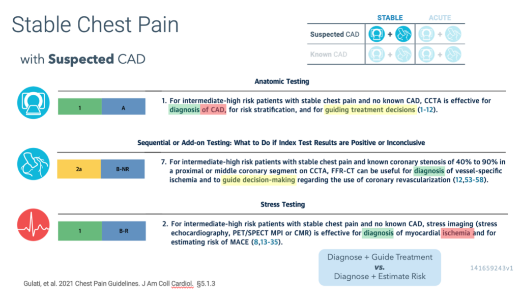
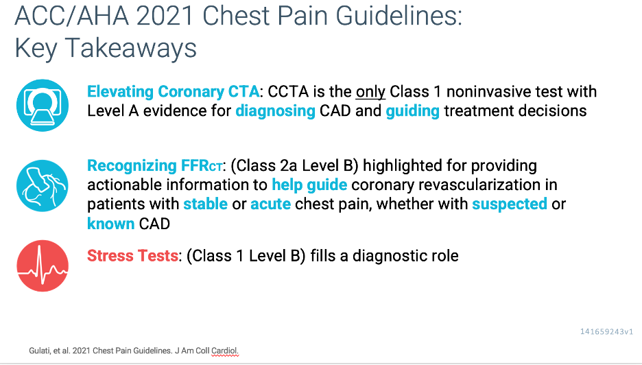
Coronary CTA as a class I Level of Evidence A for diagnosing and treatment guidance of CAD in intermediate and high-risk patients with no known CAD.

Although it is well known that guideline-based recommendations are evidence based, the evidence used to support the use of cardiac imaging has traditionally been limited, as prior data evaluating the clinical effectiveness of imaging have mostly focused on diagnostic accuracy rather than on the impact of testing on patient management and outcomes.

However, over the past decade, the quantity and quality of evidence supporting various clinical indications for noninvasive testing have grown substantially, especially for new imaging techniques.

Consequently, the new guideline was based on a comprehensive and critical evaluation of the contemporary evidence base in imaging. Priority was given to randomized controlled clinical trials that evaluated patient outcomes.

The guidelines state that “for intermediate risk patients with acute chest pain and no known CAD who are eligible for cardiac testing, either exercise electrocardiography, stress echocardiography, stress PET/SPECT MPI, or stress CMR is useful for the diagnosis of myocardial ischemia” (Class 1; LOE: B, nonrandomized), whereas “CCTA is useful for exclusion of atherosclerotic plaque and obstructive CAD” (Class 1; LOE A). Among patients with acute chest pain, invasive angiography is recommended (Class 1; LOE C, expert opinion) in the presence of moderate to severe ischemia on a current or prior (≤1 year) stress test, or in the presence of high-risk CAD (left main ≥50% or 3-vessel ≥70% stenosis), or obstructive CAD (≥50%) and frequent angina. Invasive angiography is also suggested as an option if there is moderate to severe ischemia on an imaging stress test or abnormal fractional flow reserve–computed tomography (FFR-CT) imaging after coronary computed tomography angiography (CTA) with inconclusive stenosis or obstructive CAD.

Implications for Imagers

Imagers and clinicians need to be selective in deciding who requires further testing and avoid testing in those who are unlikely to benefit from additional investigations. Intermediate-risk patients may benefit from further noninvasive testing. Similarly, as described earlier, CAC testing in low-risk individuals and a contemporary pretest probability model of obstructive CAD can be used to identify stable patients in whom testing may be deferred. In fact, when evaluating the pretest probability of obstructive CAD table (Figure 11 in the guideline), all women aged <60 years and all men aged <40 years would be expected to have a pretest probability <15%. When testing is believed to be required in such low-risk patients, calcium scoring or exercise treadmill testing (both Class 2a) may be options for the index evaluation.

Imagers also need to promote effective use of testing by ensuring that unnecessary test layering is avoided. Clinicians should use the initial test results and severity and frequency of symptoms to intensify guideline-directed medical therapies and guide the need for follow-up testing.

Imagers need to be familiar with the strengths and limitations of different imaging approaches to ensure appropriate selection of the best test. In keeping with this principle, it is important to recognize the advantages of newer testing options (eg, plaque detection or FFR-CT imaging on coronary CTA [3], higher diagnostic accuracy and quantitative assessment of myocardial blood flow with PET, use of CMR for suspected myocardial infarction with nonobstructive coronary arteries). The recommendations of this and other guidelines are based on the available scientific data, rather than on local availability or economic factors. It is thus expected that evidence-based recommendations should ultimately drive clinical practice patterns and payment, rather than the other way around.

FFR-CT imaging is suggested as a testing option following coronary CTA in patients with both stable and acute chest pain when there is 40% to 90% stenosis in a proximal or mid-vessel (Class 2a). Furthermore, coronary CTA and stress test options are presented for evaluating patients with various manifestations of nonobstructive plaque, a clinical entity that is increasingly common.

stable chest pain CAD
Acc-AHA 2021 chest pain guidelines

Sign up

to stay updated on news from pulse

Newsletter - Article

This field is for validation purposes and should be left unchanged.

Featured Articles

Start by reviewing: CCTA Reimbursement Updates for 2025 below and align your charges as seen in 1, 2, 3.

U.S. CMS doubles reimbursement for cardiovascular CT services

reimbursement for cardiovascular CT services
The U.S. Centers for Medicare and Medicaid Services (CMS) has released its final rule for 2025 reimbursement: Coronary CTA (CCTA) has been elevated to a new Ambulatory Payment Classification (APC) classification, doubling reimbursement for this procedure in the hospital setting (OPPS) and increasing payment in the physician office setting (PFS) effective January 1, 2025. [1],[2]
  1. This highly anticipated update doubles the CCTA payment rate from $175 to $357.13, aligning Medicare reimbursement more appropriately with the value CCTA provides in cardiac care. This is a win for U.S. providers as well as the entire cardiac imaging community, ultimately improving patient access to this essential diagnostic tool.
  2. What should hospitals do now that CMS has approved this change?
    Hospitals should use the cardiology revenue code (0480) for CCTA services, when appropriate.
  3. New Category I CPT code issued for AI-enabled coronary plaque analysis software
The American Medical Association (AMA) has issued a new Category I CPT code for artificial intelligence (AI) based platforms that quantify coronary plaque buildup in imaging results and identify signs of coronary artery disease (CAD).[3] The update, scheduled to take effect in January 2026, covers AI offerings from multiple companies, including HeartFlow, Cleerly and Elucid. These technologies work by evaluating coronary CT angiography images and then alerting clinicians of any findings that represent CAD and high-risk features for heart attacks.

Coding: Plaque Analysis will be billed with the 4 CPT Category III codes approved for automated plaque analysis, effective late November 2024

CPT Category III codes

Announcement comes days after CMS expanded Medicare coverage for these platforms.

October has been a historic month for AI-powered coronary plaque analysis. Just days ago, the finalized a new local coverage determination (LCD) that expanded Medicare coverage for these same technologies. Four of seven Medicare Administrative Contractors (MACs) agreed to the updated policy, which went into effect on Nov. 24.

Now that you see how to improve your revenue capture in 3 simple steps, can the rest of your CCTA program performance use a boost in revenue performance through improved efficiencies?

Pulse Imaging Consultants has more ways to improve your Cardiac CTA program, improving throughput, patient and provider satisfaction, and improving overall performance. Contact us if you would like to know more.

[1] https://scct.org/news/news.asp?id=685976

[2] https://www.federalregister.gov/documents/2024/11/27/2024-25521/medicare-and-medicaid-programs-hospital-outpatient-prospective-payment-and-ambulatory-surgical

[3] https://www.ama-assn.org/system/files/sept-2024-summary-of-panel-actions.pdf

[4] https://scct.org/page/HOPPScy25?_zs=6VO6X&_zl=DemW4
Cardiac CT Site Colorado

Another successful initiation of cardiac CT program happened last week. In Colorado mountain region where patients frequently must be air transferred to the nearest cath lab, a CCTA can significantly improve care of local patients and visitors by rapid triaging of acute patients and avoiding many unnecessary transfers to Denver.

This program will serve residents by facilitating earlier diagnostic workups for chest pain and keeping patients local, decreasing the amount of travel required to care. This cardiac CT program is also equipped with some of the newest AI programs that supplement anatomical CT images with plaque analysis and CT FFR information, providing more information to support physicians with treatment planning.

How can cardiac CT support your program with early diagnosis and keeping patient care local?